What type of joint is the patellofemoral joint?
The patellofemoral joint is a diarthrodial plane joint. The joint is made up of the posterior surface of the patella and the trochlear surface of the distal femur.
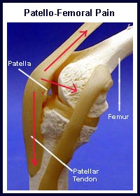
The knee cap (patella) is a small bone in the front of the knee. It is actually the largest sesamoid bone in the body. It glides up and down a groove in the thigh bone (femur) as the knee bends and straightens. The patella has a smooth coating (articular cartilage) on its underside which allows it to slide easily in this groove. The groove in the femur is called the femoral groove and it is also coated with articular cartilage. The patellar tendon is a thick, rope-like structure that connects the bottom of the patella to the top of the large shinbone (tibia).
Mikael Häggström, M.D., CC0, via Wikimedia Commons
What are the muscles of the patellofemoral joint?
The powerful muscles on the front of the thigh, the quadriceps muscles, straighten the knee by pulling at the patellar tendon via the patella. One of the quadriceps muscles, the vastus medialis, pulls the patella inward (medially). Another quadriceps muscle, the vastus lateralis, pulls the patella outward (laterally). There are also smaller rope-like structures (ligaments) on the inner (medial) and outer (lateral) sides of the patella. These small ligaments work with the quadriceps muscles to help keep the patella in the centre of the femoral groove.