This section covers basic knee anatomy. Please review this section before reading about a specific knee topic. To better understand the various knee topics covered in this website it is important to understand the anatomy and function of the knee. You may also want to review the more specific section on the basic anatomy of the patellofemoral joint.
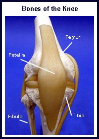
What are the bones of the knee?
The knee is the largest joint in the body and it is also one of the most complex. The knee joint is made up of four bones, which are connected by muscles, ligaments, and tendons. The femur is the large bone in the thigh. The tibia is the large shin bone. The fibula is the smaller shin bone, located next to the tibia. The patella, otherwise known as the knee cap, is the small bone in the front of the knee. It slides up and down in a groove in the femur (the femoral groove) as the knee bends and straightens. Read more about the anatomy of the patellofemoral joint.
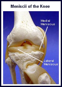
What is the meniscus / cartilage of the knee?
Two structures called menisci sit between the femur and the tibia. These structures act as “cushions” or “shock absorbers”. They also help provide stability to the knee. There is a medial meniscus and a lateral meniscus. When either meniscus is damaged it is often referred to as a “torn cartilage”. There is another type of cartilage in the knee called articular cartilage. This cartilage is a smooth shiny material that covers the bones in the knee joint. There is articular cartilage anywhere that two bony surfaces come into contact with each other. In the knee, articular cartilage covers the ends of the femur, the femoral groove, the top of the tibia and the underside of the patella. Articular cartilage allows the knee bones to move easily as the knee bends and straightens.
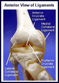
What are the ligaments of the knee?
Ligaments are like strong ropes that help connect bones and provide stability to joints. In the knee, there are four main ligaments. On the inner (medial) aspect of the knee is the medial collateral ligament (MCL) and on the outer (lateral) aspect of the knee is the lateral collateral ligament (LCL). The other two main ligaments are found in the center of the knee. These paired ligaments are called the anterior cruciate ligament (ACL) and the posterior cruciate ligament (PCL). They are called cruciate ligaments because the ACL “crosses” in front of the PCL. Smaller ligaments help hold the patella in the center of the femoral groove.
What are the muscles and tendons of the knee?
Tendons connect muscles to bone. The strong quadriceps muscles on the front of the thigh attach to the top of the patella via the quadriceps tendon. This tendon covers the patella and continues down to form the “rope-like” patellar tendon. The patellar tendon in turn, attaches to the front of the tibia. The hamstring muscles on the back of the thigh attach to the tibia at the back of the knee. The quadriceps muscles are the main muscles that straighten the knee. The hamstring muscles are the main muscles that bend the knee.
What are the bursae of the knee?
Finally, a bursa (pl. bursae) is a small fluid filled sac that decreases the friction between two tissues. Bursae also protect bony structures. There are many different bursae around the knee but the one that is most commonly injured is the bursa in front of the patella, the prepatellar bursa. Normally, a bursa has very little fluid in it but if it becomes irritated it can fill with fluid and become very large.
As mentioned above, this section covers basic knee anatomy. To read more about the anatomy of the knee please visit the links section. Links have been provided to other websites as well as online medical journals. Knee injury topics can also be accessed.